As in Incident command...

IC established!
We'll have several different sections reporting in - recent research, local topics, or highlighting areas of the Sponsor Hospital Council of Greater Bridgeport protocols.

*** Keep in mind - this website does not replace your protocols, and these posts do not reflect SHCGB or Bridgeport Hospital policies. This is a place to discuss research, controversies, or discuss possible future protocols. When in doubt, check your current protocols through the official source.

Tuesday, October 15, 2013

Fever - does EMS need to treat it?

Treating a kid with a fever seems like a no-brainer. It almost doesn’t seem to need any further justification - a fever is bad, and medicine to treat the fever is good. It’s right up there with mom and apple pie!

Did I say "no-brainer?" My bad! (source)
It may appear like we're always taking drugs (e.g. atropine, furosemide) and devices (MAST pants, pediatric intubation) away from EMS,   So, it seems pretty nifty to actually add a drug to the EMS formulary. 

And why not? The medicines (Tylenol and ibuprofen) are relatively safe (even zombie moms are allowed to buy it for their babies), EMS can start the anti-fever therapy quickly, and we can prevent….  something. 

Wait a minute, what are we actually treating?  And why is treating a fever so darn important that EMS should do it?


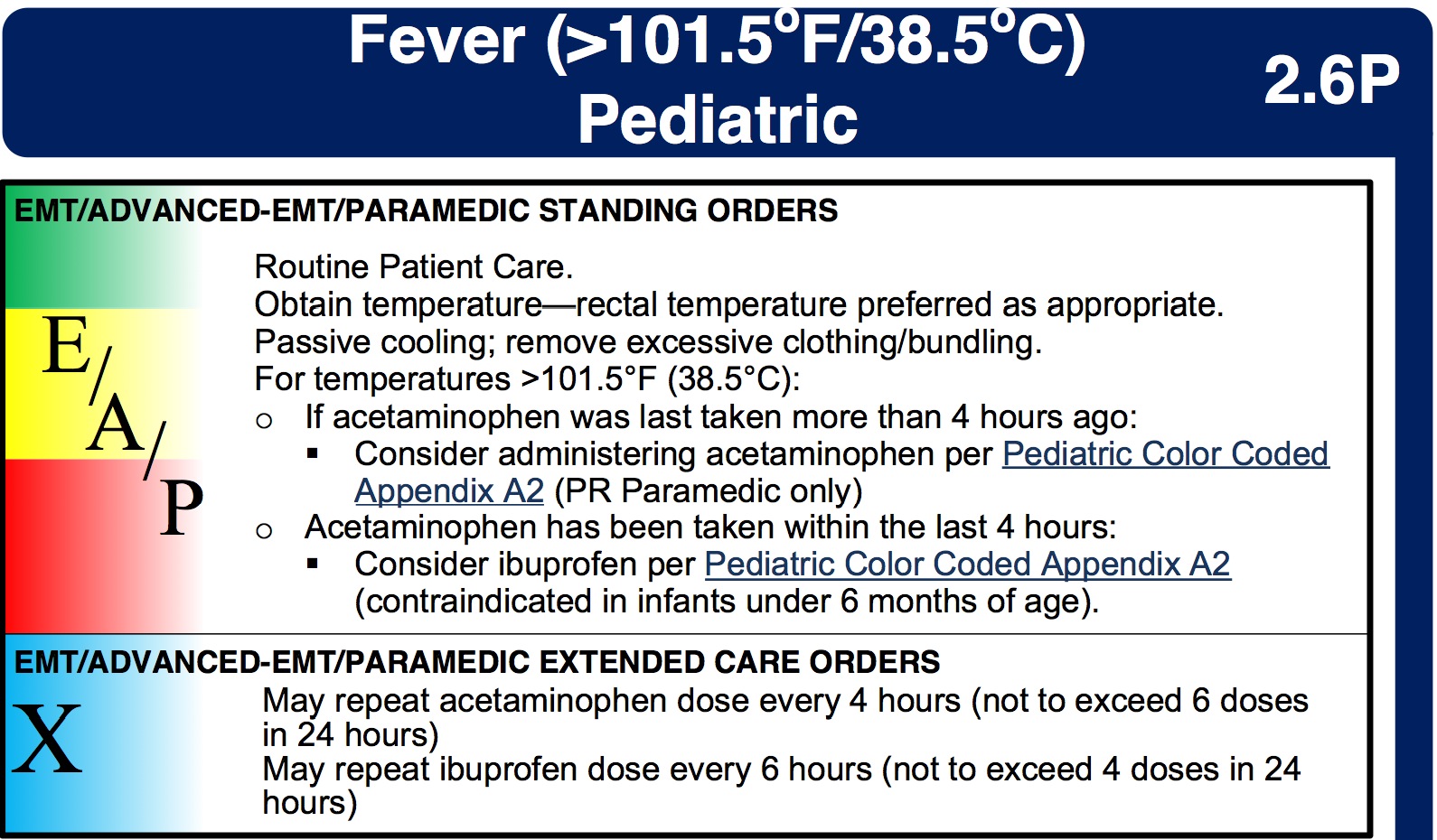
1. There is no agreement on when to "treat a fever."
Tylenol and ibuprofen are used to reduce fever, and to treat discomfort, and are near-universal therapies for kids and adults.


Treating mild discomfort is a great idea, since not all pain needs morphine. But protocols, in general, only mention a number (temperature) as a trigger for antipyretic use, and most protocols can’t even agree on which number that should be. You can find examples of fever "triggers" for antipyretic use at 100°, 100.2°, 100.4°, or even 101.5°

For example, check out the New Hampshire state protocols:


It turns out there’s a good reason why there is no agreement on what temperature "needs" APAP - nobody knows! 

And not only is there no evidence saying what temperature elevation needs antipyretic therapy...


2. Fever isn’t dangerous!
While having a fever is unpleasant, there is no evidence that it is harmful. In fact, most experts agree that damage can’t occur until the temperature has been over 106° for a while, which is almost unheard of for an infectious cause.


(Hyperthermia - as in heat stroke - is a entirely different matter, and brain damage can occur quickly, even below 106°. But no one talks about giving Tylenol to a kid that’s been locked up in a car during summer.)


The American Academy of Pediatrics makes it pretty clear 
There is no evidence that reducing fever reduces morbidity or mortality from a febrile illness.
Yeah, we should treat pneumonia or meningitis with antibiotics (or heatstroke with ice), but there is no benefit to Tylenol besides feeling a little better.


3. It won’t stop a febrile seizure, or keep it from happening again.
Yes, febrile seizures are often unsettling to parents, but they are essentially harmless. Per the experts:
There is a theoretical risk of a child dying during a simple febrile seizure as a result of documented injury, aspiration, or cardiac arrhythmia, but to the committee's knowledge, it has never been reported
But OK, they aren't, like, medically bad, sure. But why not quickly treat the fever in a kid who just seized, or who has a history of febrile seizures and just spiked a temp? Well, a number of studies have been done on the subject, and they all say the same thing - aggressive use of Tylenol or Advil does nothing to prevent the next febrile seizure. Phenobarbital or valium do prevent them, but they have significant side effects.

As the AAP guideline on febrile seizures notes that (my emphasis):

In situations in which parental anxiety associated with febrile seizures is severe, intermittent oral diazepam at the onset of febrile illness may be effective in preventing recurrence.

The paramedic’s job should be to reassure (when appropriate) the parents, and treat any mild discomfort associate with a fever. Wrestling a kid to give them Tylenol “to get their fever down” isn’t worth it, and likely doesn’t do much to help their mild discomfort. 

I'm also not sure how much the wrestling helps  treat the "severe parental anxiety!"


4. In adults with septic shock and fever, antipyretics could increase mortality
A recent issue of Chest (a journal for pulmonary/critical-care physicians) featured a spirited debate on whether septic patients with a fever should be cooled.


The only point on which everyone agreed was that antipyretics like Tylenol or ibuprofen weren’t useful, and could in fact make the situation worse. As the "pro-cooling" team in the debate conceded, 
Little evidence-based support exists for use of antipyretic medications to improve fever-associated morbidity and/or mortality.”


5. It may promote “fever phobia” in parents and medical providers.
By carrying the drug, and promoting its early use, EMS may play a role in driving "fever phobia." This is the name for the belief that many parents (and far too many doctors and nurses!) have, that fevers are very dangerous

As the expert pediatricians at the AAP put it, too many doctors and nurses worry about fever causing seizures, brain damage, or death. They then pass on these irrational concerns to the public. As they put it:
It is argued that by creating undue concern over these presumed risks of fever, for which there is no clearly established relationship, physicians are promoting an exaggerated desire in parents to achieve normothermia by aggressively treating fever in their children.
As a consequence, our medical offices, emergency departments, and EMS systems get many calls for a "kid with a fever," who doesn't otherwise look sick, wasting time and money, sometimes prompting unnecessary tests, and in general causing a whole bunch of bother.

The Bottom line
Giving Tylenol or Motrin, in accordance with your protocols, is fine. These are very safe drugs, and they do well at treating small aches and discomforts. But keep a realistic view of what you are using them for. And try to avoid spreading any medical myths!

Thursday, September 5, 2013

One more word on needling the chest.

Of course, no sooner had I posted 4 things to know before you needle the chest, when I found a new article from Australia, just published, describing how new EMS guidelines and education improved their approach to tension pneumothorax. It describes a "new" approach to tension PTX, but also has some of the limitations of the older studies. 

"Improvement in the prehospital recognition of tension pneumothorax: The effect of a change to paramedic guidelines and education."

The authors of this study out of Melbourne had been concerned by the number of patients with a unrecognized tension PTX being brought into their hospital by EMS. Their guidelines for needle decompression evidently placed a certain amount of emphasis on certain physical signs for the diagnosis of progression to tension; e.g. tracheal tugging, subcutaneous emphysema, and JVD, amongst others. 


Realizing that these indications for decompression were vague and difficult to use in the field, they re-wrote the guidelines with an emphasis on the clinical situation most likely to present with a tension - an intubated patient with chest trauma.

On top of this, they made 7.5 cm IV needles available, as well as a 10 cm-long commercial device:

This Cook device comes with a crossguard, for effective parrying.
Fewer unrecognized tension PTXs
So the new guidelines and education worked. The rate of tension PTX that was treated by EMS went from about 66% to 90%. Put another way, the number of unrecognized tension pneumos went from 10 in one year, to 4

Since the EMS service had also started doing RSI intubations for trauma, the absolute number of tension PTXs also went up - this one EMS agency needled 81 patients in one year! (BTW, what the heck is going on in Melbourne?! That's a lot of serious trauma.)

Imagine what they could do if they had guns!

The limitations
So, how do we know that the paramedics were sticking needles into true tension pneumothoraces? 

Short answer: we don't, not for sure.

Longer answer: This has been the problem with older studies that merely reported, for example, a rate of needle decompression by medics, with no attempt at verification.  One prior study, however, used the subsequent presence of an air leak from a chest tube as a surrogate for tension PTX, and found a very low rate of "true" tension PTX - just 14% of the patients that were "needled" by EMS. An ultrasound-based study also casts doubt on the prehospital diagnosis of tension PTX.

The authors of the current paper used a "case definition" to identify tension PTX. That is, they went over all the clinical records and imaging studies, and made a best guess about what the patient had in the field. As you might imagine, this is an imperfect method, and we can only guess at the accuracy of the paramedics' diagnoses.


The bottom line
Despite these concerns about accuracy, there were fewer patients brought into the ED with a tension PTX, even as the rate of RSI, and thus positive-pressure ventilation, increased over the study period. That's a good thing, and it seems as though there was no large increase in complications. 

In the meantime, if anyone is planning a large prehospital study to look at PTXs, it sounds like Melbourne is the place for you!


Saturday, August 31, 2013

4 things to know before you needle the chest.

Looking for a tension pneumothorax (PTX), and especially deciding to perform a needle decompression, can trip up even the most smartest EMS provider. There's a balance between the need for aggressiveness, and the wisdom of careful monitoring.


Ok, that was too aggressive - dial it back next time.
A few recent cases brought into Bridgeport Hospital, as well as some recent research, help us refine the approach to evaluation and treatment. 4 fun facts about tension PTX!

1. It's rare!
While some authors have described a 6% rate of tension PTX in prehospital trauma patients, the rate is probably far lower in most U.S. EMS systems, with some estimating it happens in fewer than 0.3% of blunt trauma patients.

Another study, published in 2013, found that about 1% of the trauma patients coming into a Level 1 center had received a needle by EMS for a suspected PTX, and that most of them ended up getting a chest tube in the ED or OR. 

Problem is, they weren't able to tell if the patients had had a tension PTX when EMS needled them, or just a plain ol' pneumo. So, at worst, 1% of severe trauma patients have a tension PTX.

2. Paramedics have trouble identifying it.
That last study showed that  paramedics are pretty good at diagnosing a pneumothorax. How good are they at diagnosing the tension part?


A study done in Nashiville looked at 19 patients who got a needle decompression by EMS for suspected tension. About 1/4 of those patients were proven to have any sort PTX (tension or not), and only 2/14 of the living patients were considered to have had a tension PTX (based on finding an air leak after placing a chest tube).
So, only 14% of patients who got a needle actually had a tension PTX

3. Diminished lung sounds & crepitus ≠ tension
A patient can have complete collapse of a lung, and still not need a needle!

A few months ago one of the medics was concerned that they hadn't treated a trauma patient as aggressively as he should have. The patient had fallen a few days prior, and was complaing of shortness of breath. The medic noted bruising over the chest, paradoxical chest wall motion, and absent lung sounds. Nevertheless, the patient was breathing better on 2 lpm of oxygen, and the vitals were fine.

In the ED, the patient was promptly sent to the CT scan...
Pneumthorax, as well as subcutaneous air
... and got a chest tube right away! So the medic was concerned - should they have placed a needle in the field?

It doesn't sound like it. It's important to remember why a tension pneumo is bad. Progressive collapse of the lung leads to hypoxia, and the rise in intrapleural pressures compress the IVC, leading to hypotension. In other words, patients don't die from a pneumothorax - they die from hypoxia and shock. Rogue Medic wrote about this at some length, and you might enjoy his take on the subject.

So if subcutaneous emphysema and absent lung sounds don't require you to decompress, what signs should? A very thorough (and free!) review article from 2005 provided a list of the signs that trauma experts agree could warrant EMS attempting to decompress.

Just remember - hypoxia and hypotension!

Source
(Keep in mind that you should be suspecting tension pneumothorax - don't needle the patient with bradypnea, pinpoint pupils, and track marks just because she's hypoxic with a decrease LOC.)

It's only helped in one case report.

4. Your needle may be too short (or your patient too big...)
So, another medic brought in a trauma patient, thrown from a motorcycle, with pain over the left chest, and absent lung sounds as well. The medic had already needled the chest with the standard 14g IV catheter, but reported that lung sounds were still absent.

Sounded like a no-brainer to me; if the patient hadn't already needed a chest tube before, now they most likely did (since a needle decompression can also cause a pneumothorax). But when the patient was rolled into the trauma bay, we found no pneumo on chest x-ray, ultrasound, or even CT. Heck, looking at the CT, we realized there was no way the stubby 14 g ever made it near the lung!

This is a very common experience - unless you have a special long catheter, you may not be able to reach the pleural space, especially in the standard "midclavicular, 2nd intercostal space" that we've been taught. Why? Because we're all getting bigger, and the catheters are not!

A standard 14g iv catheter; 2" or 5 cm,

Seems big at first, but take a look at where we're trying to put this needle:

People can be thick, both in the 2nd ICS anteriorly (left),
as well as the 5th ICS laterally (right)
(Adapted from Schroeder et al)
That can be a lot of skin, fat and muscle to get through! A standard 5 cm catheter may reach the pleura in most Japanese patients, and some Turkish patients, but only in a minority of Americans.

One option is to place the needle at the 5th ICS, along the anterior axillary line, since there is generally less muscle and fat there than at the traditional 2nd ICS. One study, done in Los Angeles, found that a standard 5 cm (2 inch) needle placed at the 2nd ICS would be too short in 42% of patients, but only in 15% at the 5th ICS.

So, a lateral placement might help, but so might more needle. They make a longer 14 g iv catheter just for decompressing, extra long at 3.25 inches, or 8 cm. Of course, what's the downside of having an extra-long needle shoved into the left side of the chest?

"With a PTX, is the blood supposed to shoot out?"
The Bottom Line
A pneumothorax happens relatively often in severe trauma, but these do not require any specific prehospital treatment. A tension PTX, on the other hand, is rare, difficult to diagnose in the field, and there are some obstacles to treating it appropriately.

You're looking for a patient with not just absent lung sounds, but persistent hypoxia, and worsening hypotension. And when you place the catheter, you have to make sure it's actually reaching the pleura - but not too far in!


Wednesday, July 10, 2013

Medics for cath lab activation of STEMI? Non merci!

Although we often talk about "prehospital activation" of the cath lab for STEMI, it isn't always clear what we're talking about. For example, in some EMS systems, the medics give a "heads-up" to the ED, so that a physician can meet them on arrival to confirm a STEMI. Other systems rely on the paramedics to interpret the ECGs themselves, and make a judgement about activation on their own initiative.

A group of Québécois cardiologists, however, decided that they wanted to try a new system, where neither the paramedic, nor the ED physician, would make the decision. Heck, it wouldn't even be a cardiologist. Instead, the turned the decision over to...


"Beep boop click tweet" (Translate: "I could go for some poutine.")
The Study
The prehospital providers in Laval, Quebec, were trained to obtain a 12-lead ECG on patients with chest pain or dyspnea, but did not receive any instruction on interpretation. If the machine (a Zoll, not actually an R2 unit!) gave an interpretation of *** AMI ***, the EMTs put in a call to the hospital paging system to activate the cath team. The ECG was not immediately analyzed or transmitted, although it was saved.

Over the course of 2 years the EMTs activated the cath lab 157 times, and most of those went to angiography and were found to have coronary occlusion (a "true STEMI"). A few patients (5%) had a very suggestive ECG, but had nothing on cath ("false positive activation"). It happens.


Unfortunately, 12% of the group were found to be "inappropriate activations," meaning that the ECG shouldn't have led to activation. The authors divide these up into human factors (poor quality tracings, lots of artifact), and machine factors (the machine tried to interpret STEMI during a SVT).

What's helpful about this study
It's good that they have distinguished between "false-positive" and "inappropriate" activations, since these concepts often are mixed up in these kinds of studies. They also highlight the importance of obtaining a quality ECG, free of artifact, as well as the perils of diagnosing a STEMI during tachycardia.

What's not so helpful
The authors focus quite a bit on decreasing the rate of non-STEMI cath lab activations, which is of course a worthy goal. But there is little discussion of the STEMIs that EMS did not catch with the automated system. I.e., this study demonstrated the specificity of the system, but said nothing about the sensitivity.

Look at it this way. Laval is a big town, with about 400,000 residents in the city. Based on estimates of the incidence rate of STEMIs (about 100 STEMIs/year per 100,000 people), there should have been about 800 STEMI patients in Laval during the study period.

In other words, where were the other 800 - 128 = 672 STEMIs? 

How many of those 672 patients were transported by EMS? How many were "missed," either because of a bad-quality tracing, a misinterpretation by the algorithm, or because they presented with atypical symptoms, and so never even  received an ECG by EMS? We have no idea.

So, while decreasing false or inappropriate cath lab activations is a worthy goal, so is making sure that we're not missing anyone who should be sent there!

On the other hand, in Denmark...
A recent article out of Denmark described the experience with a "modern" approach to cath lab activation (e.g., activation based on smaller degrees of ST segment elevation, inclusion of old LBBB patients, use of EMS activation). The Danish cardiologists found a whooping 20% rate of negative coronary caths - and were totally fine with it. In their discussion, they express more concern with missing the opportunity for an early intervention, than with the "false negative"rate. They conclude that
a triage program with acute angiogram for all suspected STEMI patients should anticipate that 1 of 5 patients will not need primary PCI.
Now, I'm not endorsing this rate of negative caths as a goal, and some of their comments suggest that they may have swung the pendulum a bit too vigorously (E.g. "liberal access to acute coronary angiography means some of the patients had a “rule-out” angiogram, for example younger patients with chest pain and an ECG compatible with pericarditis.").
Nonetheless, it's clear that the authors are concerned with catching everyone they can in their cardiology "net."

So, should we take medics out of the loop on STEMI?
Well, as usual, "it depends." There are so many local factors that go into the design of every EMS system, and this may be the most appropriate response, say, in an area that will likely never be able to support the training and QA/QI that ALS systems require. And, frankly, if all you want your EMS system to do is identify the obvious STEMI cases, this is probably the way to go.

Except, you probably want your EMS system to do more. 

You want to identify and treat the NSTEMI patients who need early intervention, as well as STEMI patients. You want medics to identify the potential, "hyperacute," STEMIs. You want them to find and treat CHF, distinguishing "cardiac asthma" from plain ol' asthma, and even discriminate between primary and compensatory tachycardias.

But the "automated" STEMI activation doesn't provide any benefit beyond getting one small slice of the EMS patient population earlier care. That's fine, great even, but it's still a limited benefit. Why not aim just a bit higher?

Friday, July 5, 2013

"Cardiac anaphylaxis" after IM epinephrine?

Can IM epinephrine, at the proper dose, cause an MI? A lot of paramedics worry about this, and, as a result, under-treat anaphylaxis. This is a problem, since this is one of the small group of problems where paramedics can truly save a life.

Another example: whatever this guy is doing (source)
Of course, some of the previous descriptions of complications after epinephrine therapy involve mistakes in dosing or route. For example, this case report states that a young woman developed an AMI after getting "low-dose" epinephrine. Well, she actually received 100 µg IV, or 0.1 mg, which is about ten times what these allergy experts used in their study

"Start at 5-15 µg/min"
NOT 100µg/min.
Or even worse - how about giving 2mg epi IV instead of Narcan! Can you imagine the paperwork you would have to fill out after doing that?


A recent case report might cause some EMS providers to worry about administering the appropriate IM dose of 0.3 mg. I don't think that it should - let me explain more.

"Cardiac anaphylaxis: A case of acute ST-segment elevation myocardial infarction after IM epinephrine for anaphylactic shock."

This case report describes the clinical course of a middle-aged gentleman:

A 62-year-old male smoker with no other comorbidities presented to emergency department at 6 am with complaints of generalized pruritus and shortness of breath after taking diclofenac for toothache 1 hour back. On examination, pulse was 97/min; blood pressure, 84/60 mm Hg; jvp, normal; cardiovascular system, unremarkable; respiratory system, rhonchi bilaterally.
Sounds like anaphylaxis! The  ECG, before epinephrine, showed:

"Nonspecific [ECG] changes on arrival"
He then received 1 mg epinephrine IM, which is 3 times the recommended dose. Interestingly, they describe the IM administration as having been given over 5 minutes.

A second ECG was obtained after the patient developed chest pain:

He underwent PCI, and a thrombus was sucked out of his LAD. He ended up doing well.

Was the STEMI really due to the epinephrine?
I'm not so sure - take a closer look at the initial, "non-specific," ECG:
Close up of V1-V3
That looks like quite a bit of ST elevation, especially relative to the QRS, in V2 and V3. But why would someone have a STEMI before getting epinephrine?

Well, sometimes anaphylaxis itself can cause an MI. It's called Kounis Syndrome, and there a number of case reports out there:

Acute coronary syndrome triggered by honeybee sting: a case report.

ST-segment elevation myocardial infarction following a hymenoptera (bee) sting.

Acute anterior myocardial infarction after multiple bee stings.

The Bottom Line
The authors acknowledge this possibility, and also acknowledge that epinephrine-related MI is not typical.
Acute myocardial infarction (MI) following anaphylaxis ("cardiac anaphylaxis") is rare. Epinephrine causing ST elevation in these anaphylactic patients is even more rare.
 In this case, I wonder about the initial "nonspecific" ECG, and the role of epinephrine in causing his STEMI.  Despite the authors' certainty that "high-dose epinephrine 1 mg (1:1000) IM has triggered the formation of a thrombus in the left anterior descending artery," I wonder if the STEMI was underway before they gave the epinephrine. 

What do you think?

Monday, June 17, 2013

Atrial Fibrillation: Sure, you can cardiovert it! But should you? (Part 2)


  In the first half of this post I emphasized a few points about AF. It can be triggered by a variety of non-cardiac sources - hypovolemia, alcohol, and especially sepsis can all exacerbate chronic AF, or provoke a new episode. You can cardiovert a hypotensive AF patient with any of those problems, and it might (might) fix the rhythm, but it would be unlikely to correct the underlying issue.

Need another example? Alright, how about AF and an ECG that suggests STEMI?
 
AF and proven STEMIs - first case
The examples above weren't actually hypotensive, but this last patient was. 

An elderly patient with a history of paroxysmal AF was transported by EMS for acute onset dyspnea, chest and abdominal pain. Things got more complicated when she arrested right upon arrival in the ED. 

Not the patient - this is me when the patient arrests 10 seconds after arrival.
 Fortunately, this meant that the patient got full, immediate, and successful resuscitation. The post-ROSC ECG, however, was concerning:


The patient had a pressure of 80/50 - what should the team have done next? If you say cardioversion, what would you have done if the first attempt didn't work? (or the second?)

(Also, does it help if you know that the prehospital ECG looked like this?)

Anything jumping out at you?
Well, you need a plan B in atrial fibrillation! In this case, the team decided not to re-shock a patient who had just regained their pulse, and who was apparently undergoing a STEMI. During emergent cardiac cath, a complete occlusion of their left anterior descending artery was found, and successfully stented.
 
AF and proven STEMIs - second case
I've talked about this case before, so I'll be brief. Midlle-aged woman, acute onset chest symptoms:


However, once the medic brought the rapid ventricular response (RVR) down a bit, and the symptoms improved a little, the computer message disappeared when the repeat ECG was obtained...


... but not the ST elevations, nor their apparent reciprocal changes. Her old ECG showed very normal inferior ST segments, supporting the diagnosis of an acute STEMI. During the emergent PCI, they found that an old stent in her RCA was 100% occluded.

On the other hand...
Some physicians are fairly skeptical about ST changes that are found in AF with RVR. As with other arrhythmias, you can end up with a variety of ST changes that resolve with the tachycardia. PSVT very commonly produces ST depressions, even in young folks with no heart disease. 

For example, despite the dramatic ST depressions (and aVR elevation!), this patient... 
ECG from a great case at EMS 12-Lead
 ... never had a troponin increase - no MI - and the ST segments normalized after cardioversion.

Stephen Smith, of Dr. Smith's ECG blog, also voices wariness about calling a STEMI in AF with RVR. He has a great case at his site that illustrates the lesson that, if the patient is losing units of blood from their GI tract, the cath lab is probably not the best first stop, even if the ECG computer is trying to tell you otherwise!

Pictured: Not a cath lab candidate.
So, when do you activate the cath lab? Do you wait to make the call until you've loaded the patient with diltiazem, or do you do it first thing? How long do you look for other causes, versus get everyone moving to the lab?

Ah, good question, and I wish I had some hard and fast answers. I don't know of any research that looks at this issue, and the experts can disagree. It's often going to depend on the clinical context, as well as evolutions in the ECG findings, echocardiograms, and comparisons with old ECGs - all of which are hard to do in the back of a rig!

The Bottom Line
This is a cornerstone of emergency medicine - if the rhythm is fast, and the patient is not doing well, and you think they are not doing well because of that rhythm, then the patient should be cardioverted. 

(Repeat - "they are not doing well because of that rhythm..." Important!)

This is clearly supported in our SHCGB protocols:


But we have seen in these posts a number of examples where cardioversion probably wouldn't have been effective, since the underlying medical issues needed treatment. Cardioversion isn't going to treat low magnesium, hypovolemia, and especially not sepsis!


Cardioversion for hemodynamically unstable AF is reasonable, but this isn't as "simple" as ventricular fibrillation. You need to consider the causes and aggressively treat them, and be ready with a "plan B."