As in Incident command...

IC established!
We'll have several different sections reporting in - recent research, local topics, or highlighting areas of the Sponsor Hospital Council of Greater Bridgeport protocols.

*** Keep in mind - this website does not replace your protocols, and these posts do not reflect SHCGB or Bridgeport Hospital policies. This is a place to discuss research, controversies, or discuss possible future protocols. When in doubt, check your current protocols through the official source.

Tuesday, February 18, 2014

Sudden Cardiac Death Among Firefighters ≤45 Years of Age

I want to talk about a new study that looks at heart attacks in younger firefighters, so no jokes or funny picture today. It’s hard to come up with a humorous spin for a graph like this: 
From a 2012 FEMA report
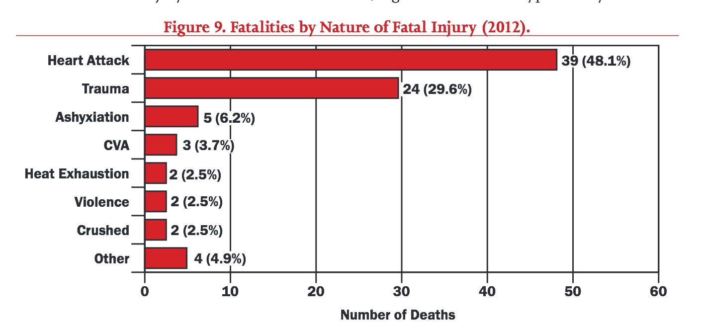
The chart above comes from the 2012 USFA report of firefighter fatalities, and it’s shocking. Over half the fatalities in that year had nothing to do with burns, entrapment, falls from ladders, or other traumatic dangers. Instead, MI and stroke, in general, were the biggest danger firefighters faced. 
Now, this is partly a function of age, but 15% of the deaths from MI were in firefighters younger than 45 years old. A recent study focused on this group, and found some surprising results.
The Study
The authors of Sudden Cardiac Death Among Firefighters ≤45 Years of Age in the United States wanted to take a closer look at these younger firefighters who died of cardiac causes, and see if there were any risk factors that could explain those deaths. 


To do this, they used the database maintained by NIOSH (http://www.cdc.gov/niosh/fire/) to find all cases of firefighters < 45 years of age who had a sudden cardiac death. They looked at the period from 1996 to 2012, and examined autopsies and other reports.
They then selected a bunch of age-matched, healthy, “occupationally active” control firefighters to compare them to. Additionally, they looked at noncardiac traumatic fatalities (deaths due to blunt trauma, burns, or asphyxiation) to serve as a second comparison group.

1. The FFs with sudden cardiac death 
They found 87 FFs under the age of 45 who had a sudden cardiac death during that time. 

A few results stand out:
  • Almost all were men.
  • Over 1/4 of them smoked 
  • Almost 2/3 (63%) of them had a BMI ≥ 30
  • Over 1/4 of them had a BMI over 35
  • Over 1/2 had evidence of both cardiac disease and cardiomegaly
At first glance, it looks like these firefighters had much worse health than the general public. But maybe this is due to the unique stresses of the job. For example, disruption of normal sleep patterns could encourage sleep apnea, leading to hypertension and obesity.

So in order to clarify the issue, they compared the FFs who had sudden cardiac death with the people most like them - other firefighters!

2. Compared with occupationally active FFs
When they compared these FFs to the occupationally active" FFs, however, they found concerning results, suggesting that the FFs who had cardiac arrests were indeed unhealthier than their own peers.


Obesity, smoking, and hypertension were significant predictors of cardiac death while at work. Not unexpectedly, a history of cardiac problems was a huge risk factor as well.

3. Compared with FFs who had a traumatic death
When they then compared the FFs who died from sudden cardiac death to those that had a traumatic death, they found significant difference in both age, and in the size of their hearts. FFs who suffered a cardiac death had higher rates of cardiomegaly, or enlarged hearts, suggesting that they had had longstanding problems with hypertension or obesity (or both).


So what do we do with these results?
First off, prevention (i.e. lifestyle) trumps everything. Quitting smoking, keeping a healthy weight, and maintaining a vigorous exercise routine, amongst other things, may have gone a long way towards preventing many of these deaths. Although firefighting presents many unique challenges to staying healthy (e.g. schedules that disrupt sleep patterns, exposure to heat stress), the comparisons to healthy FFs, as well as those who died from trauma, show that smoking and hypertension play a huge role in raising the risk of cardiac death.

Second, we need to prepare for cardiac arrest in firefighters. The unique nature of the fireground, as well as the obstacles that the clothing and equipment present, mean that departments need to practice their response to a "fallen" firefighter, aiming to start CPR and assess for a shockable rhythm as soon as possible. This requires special procedures, teamwork and practice. Watch these guys from Hilton Head FD run through a drill.


Be safe, and take care of your heart!

Sunday, February 9, 2014

Can you give adenosine to a patient with WPW?

Maybe you can answer the question in the title completely and confidently. If so, feel free to skip this post, and go on to some more entertaining corner of the web. However, it seems to me that many emergency providers are unsure about how to approach a confusing issue with adenosine:
Can (or should) you give adenosine to a patient with known or suspected WPW?
Spoiler: The answer to this questions is

More specifically:
  1. Adenosine is safe and effective for terminating SVT (narrow or wide) caused by WPW, but...
  2. Giving adenosine for WPW with atrial fibrillation, however, can be lethal.
One recent case that was treated in the field illustrates principle #1. Fortunately, no local medics have provided us with a demonstration of principle #2!

(As always - Follow your local protocols for WPW and arrhythmias!)

Can you give adenosine to a patient with WPW?
Case report:
This is best illustrated with a case! Paramedic Harvey had a patient with palpitations, tachycardia, and a good BP, and she obtained the following ECG.


A narrow-complex, regular tachycardia in a stable patient? Looks like an SVT!  Medic Harvey decided, appropriately, to bust out the adenosine.

A rapid bolus of 6 mg broke the tachycardia, and restored NSR on the monitor, but the follow-up 12-lead looked a little odd:


A few minutes later a second post-conversion ECG was obtained.


Huh. You know, that sort of looks like...

It was at this point that the patient told Heather 
"I probably should have told you that I have WPW."

Wait, what?!
Oooooo.... Isn't WPW supposed to be a dangerous rhythm, where you can send the the patient into VF just by thinking about adenosine? Holy cow, did the medic just get lucky, or what?

Brief Review of WPW
Let me go over the pathophysiology briefly. 
(For a fuller description, check out a great new e-book by Ken Grauer, entitled ACLS - 2013 - ePub, for more education on WPW and other topics in ECG interpretation and ACLS.)

The 1993 edition was pure gold when I was in paramedic school.
It's only gotten better - seriously, download this now!
There are 2 common causes of a regular narrow-complex tachycardia, or SVT
  • The most common is AV nodal reentrant tachycardia (AVNRT), which involves a "loop" of electrical current that is confined within the AV node. These are narrow unless aberrant conduction occurs.
  • By contrast, AV reentrant tachycardia (AVRT), involves a "short circuit," or accessory pathway, between the atria and the ventricles that bypasses the AV node. During normal sinus rhythm, the ECG will often demonstrate signs of "pre-excitation" of the ventricles; a short PR, a delta wave, and a wide QRS.
Grauer K: ACLS-2013-ePub, KG/EKG Press (available in kindle/ibooks/nook/kobo)
When an episode of AVRT (aka SVT) is triggered, however, the QRS will usually be narrow, and the delta wave will disappear (as seen in Panel A below). As noted in Panel B, however, in a rare minority of patients the circuit of conduction will be "backwards," and produce a wide QRS.

Grauer K: ACLS-2013-ePub, KG/EKG Press (available in kindle/ibooks/nook/kobo)
So, there are some important differences between the two main causes of SVT (AVNRT and AVRT). It's important to emphasize, though, that...

AVNRT and AVRT have 3 important things in common:
  • Both rely on the AV node to complete the "loop" of electrical current that generates the tachycardia;
  • In both, the sinus node is generating normal, controlled signals; i.e., 60 - 100 beats per minute.; and 
  • Adenosine will terminate the reentrant thythm in both by shutting down the AV node.
In AVNRT, adenosine effectively "cuts" all electrical connections between the atria and ventricles (temporarily!). In AVRT/WPW, however, the bypass tract is unaffected by the drug, and is still capable of conducting electrical signals to the ventricles. This could potentially lead to sinus impulses being conducted anterograde down the AP, producing wide, "aberrant," QRS complexes, but at a controlled rate (i.e. 60-100 bpm).

WPW with AF can be lethal if treated with adenosine
This is entirely different when the patient has WPW and atrial fibrillation. Remember that, in AF, the atria are firing off at 300 - 400 times a minute, not the controlled rate of 60-100 that the SA node generates. 

So, if these all of these electrical impulses are transmitted to the ventricles through the bypass tract, the myocardium will freak out. Like this.

Source: Shah 2001

WPW without AF is safe to treat with adenosine
We probably don't need to worry*, however, about giving adenosine in patients with WPW, but without AF. There's a few reasons why.

1) First off all, about a third of the SVTs that you have given adenosine to in the past were actually caused by AVRT/WPW, just based on the epidemiology. Since practically none of these developed VF (right?), this is apparently a very safe practice.

2) Second, there was a concern in the past that a certain percentage of wide-complex tachycardia were actually WPW with antidromic conduction, and so the advice was to avoid adenosine. The rationale was that since the bypass tract was capable of retrograde conduction, shutting down the AV node could "expose" the ventricles to potentially unregulated pacing. However studies such as this one have convinced a number of people, including the AHA, that adenosine is pretty safe in anyone with a (regular!) wide-complex tachycardia. 

3) Third, many cardiology experts believe that adenosine is safe in regular WCTs. The authors of a 1991 study found it to be quite safe in this setting, and concluded that 
"Adenosine may cause acceleration of preexcited atrial arrhythmias, but these effects are transient and should not discourage the use of adenosine as a diagnostic agent in broad complex, regular tachycardias of uncertain origin."
Dr. Stephen Smith, of Dr Smith's ECG Blog fame, believes that we don't even need clinical evidence to prove that adenosine is safe in regular tachycardias, wide or narrow, in a person with a history of WPW. A fundamental understanding of cardiac physiology is enough to sh0w this:
"Anyone who knows what AVRT is, and what it is that electrical circuits do, knows that it is safe to give adenosine. If you ask an electrophysiologist for an article on this, they will say 'There are no articles, because this is so obvious that it needs no proof.'
[A]denosine is safe in VT. One need not prove that it is safe in AVRT ."

The Bottom Line
So it turns out that adenosine was quite safe with Harvey's patient, as well as effective. This probably wasn't a fluke!

Although many medics have been taught that Wolff-Parkinson-White (WPW) and adenosine are a dangerous combination, this isn't often the case. On the contrary - it seems likely that medics and physicians have frequently given adenosine to patients with undiagnosed WPW, without apparent ill effects. 

Furthermore, the recent emphasis on giving adenosine for regular wide-complex tachycardias of uncertain etiology makes it even more likely that YOU will give adenosine to someone with undiagnosed WPW. A small number of those WCTs represent WPW with antidromic conduction, but adenosine administration has not been documented to cause any problems to date.

So, follow your local protocols for arrhythmias and WPW, but keep in mind the available evidence, as well as expert opinion, suggests that adenosine is safe in most cases.
__________________________________
* Okay, there are a few, totally theoretic, reasons to worry about giving adenosine to patients with WPW. I mention these only out of a sense of completeness. 

First of all, adenosine is not totally benign, and it has been shown to induce atrial fibrillation in a number of case reports. Although the half-life is fairly short, it's conceivable that adenosine could first trigger AF in a patient with a history of WPW, and that the AF could then immediately degenerate into VF. One case report describes how a young woman with SVT developed AF after performing a Valsalva maneuver. Adenosine was then given, and the rhythm degenerated into a pulseless irregular WCT. Cardioversion restored a pulse and sinus conduction.

Second, although there are no case reports that I know of have shown an antidromic WPW tachycardia deteriorating after adenosine administration, this is a rare rhythm, and even a moderate relative risk of adverse effects from adenosine would produce a small absolute number of complications. For example, the study by Maril (that demonstrated the safety of using adenosine in WCTs) only enrolled 2 patients with a history of WPW. One of those patients was determined to have been having a ventricular (non-accessory pathway dependent-) rhythm upon enrollment. It's hard to determine the absolute safety of a drug for such a rare rhythm. 

Nonetheless, even a "worst-case scenario" suggests that an adverse event would be very rare.

Monday, January 13, 2014

Pediatric Anaphylaxis: Medication errors by EMS

You are called for a 5-year-old child with trouble breathing. 

The mother states the child has had 1 hour of progressive respiratory distress after being stung by a bee. The initial vitals are BP 76/40, P 120, R 45, and SpO 91% on room air. The patient is sitting upright, speaking in short sentences, and has audible wheezing. The physical exam reveals hives over the chest and arms.  If you don't act in 2 minutes, your patient will decompensate. What do you do?

(By the way, this is a HIPAA-compliant picture of your patient:)


Yup, it’s a replicant, and just like Roy Batty, he was programmed to die in this EMS simulation study performed in Michigan. 

Unlike Roy Batty, however, this replicant will live if he is given epinephrine in time!

"I've seen things you people wouldn't believe...  And I need some epi!"

The authors used a high-fidelity simulation manikin, in a well-equipped simulation center, as well as the scenario described above. They allowed the EMS crews to use their own medication and equipment, but they all had to follow the state EMS protocol for pediatric anaphylaxis. 

Michigan EMS protocols
 If the crews gave the right medications and interventions, the kid got better. If not....

So what happened? 

Before I answer that, let me point out that the EMTs and medics volunteered for this study. This suggests that these folks were motivated enough to participate in research, aware they were going to observed, and fairly confidant in their knowledge and skills. In other words, probably some good, smart people.

So, it's all the more distressing what the study found.

1. Three out of 62 crews (5%) did not give epi 
For true anaphylaxis, epinephrine is the required treatment, with essentially no contraindications. 


2. Epi was often given by the wrong route. 
The protocol spelled out that epinephrine should be given IM, in line with several national and international guidelines. Despite this, only 37 out of 59 (63%) crews gave epinephrine IM. The authors were generous, and allowed SQ as an acceptable route, but this is an outdated practice. 

3. An epi dose of > 1 mg was given by 20% of crews. 
‘Nuff said about that... 

4. Epi was given intravenously by 15% of crews 
Although IV epi is listed in the protocol, even "low" doses of IV bolus epi can cause badness. With that in mind, the protocol allowed for IV epinephrine only “in cases of profound anaphylactic shock (near cardiac arrest).” 

Because the patient was initially sitting upright, speaking, and perfusing, IV epi was considered a major error.

5. Less than half of the crews gave epi by the right dose and route. 
As shown in this table:


So why did this happen?
It's hard to manage a rare medical event, using a drug that is available in multiple concentrations, and can be given by multiple routes, but must be dosed accurately by weight.

And as I said before, these were engaged and motivated EMS providers. Furthermore, the errors that they committed have been demonstrated to occur in a number of prior studies and case reports, involving a wide range of medical personnel.

In other words, you can’t respond to this study by saying “Well, our guys would never screw up like this!” Or by saying “We’ve been doing it this way for years without a problem.” This study should force all of us to reevaluate how we teach, protocolize, and practice treatment of anaphylaxis.

If you have the patience, download the author’s summary (the picture below) of the types of errors that were committed, and the rationale of the medics who committed them. Very informative!


Friday, January 3, 2014

The Cardiac Save Pin

I’ll admit, I don’t understand why the stork pin is so popular. 

The mother is doing all the pushing and tearing, while the medic or EMT isn’t really doing any work besides not dropping the newborn. That’s not really an advanced medical skill, you have to admit.
"Good job holding a human off the floor!"
Our new “Cardiac Save” lapel pin, on the other hand, recognizes the judgement that medics use in identifying a STEMI, and then communicating effectively with the ED and cardiology. These skills aren’t as dramatic as, say, sinking an ET tube or needle decompression, but they are arguably far more important.
And they're made in New England! (site)
I wanted to illustrate this with a few recent STEMI cases. Basically, I just want to brag about EMS in Bridgeport!

Case #1 - Not an obvious STEMI
See what you think about the ECG. Older female, late at night, chest pain:


Kind of a tough one, since the RBBB mucks up the QRS. Unlike LBBB, however, the ST segments should be basically normal in RBBB. The paramedic, Gordon MacCalla, from VEMS, also thought this was a STEMI, despite the fact that the computer took a little longer coming to that conclusion than he had. 

Once in the ED, this ECG had the cardiology fellow scratching their chin, but the interventional cardiologist only needed about 0.25 seconds to verify the STEMI before heading to the cath lab with the patient. Since this all happened late at night, the cath team needed to be called in from home, so Gordon's prehospital activation saved plenty of time and myocardium!


Case #2 - Bypassing the ED
It’s not always possible for EMS to go directly to the cath lab, since the cardiac team may not have assembled by the time EMS gets to the hospital (as in case #1). Case #2 was the first time where the timing worked out, and paramedic Erin Smith, of Stratford EMS, got to skip the ED.

This middle-aged gentleman was actually hypotensive when EMS arrived. He complained of chest discomfort, and the first ECG showed:

Not subtle. Yeah, it was the RCA. By calling this in from the field and skipping the ED, medic Smith helped save this guy some heart muscle. The patient did well, spending less time admitted to the hospital than do most women after childbirth!


Case #3 - STEMI and cardiac arrest
Paramedic David Rodriguez of AMR had his hands full on this call! A not-old male had some chest discomfort, and called EMS instead of waiting it out. Good move.

Dave obtained the first ECG:


Nasty ST segment elevations in the anterior leads, eh?

That was bad enough, but then the patient went into VF! Fortunately, the time to first shock was minimal, since they were in the back of the ambulance at that point, with the defibrillator an arms-length away. After just one 200 joule shock* the patient was back in NSR, with a pretty brisk return to consciousness. At the hospital, he went immediately to the cath lab, where a proximal LAD lesion was opened.

This case is a good reminder - always think of STEMI with a cardiac arrest! Once you get a pulse and a blood pressure back, do an ECG if it hasn’t already been done.
Case #4 - Fantastic Door-to-Balloon times
Sorry, no ECG, but in a way that’s a good thing. Let me explain…


Paramedic Dane Johansson from Stratford EMS responded to a call for a middle-aged male with chest pain. On-scene he quickly performed an ECG, found a large anterior STEMI, and immediately called for a cath lab activation.


Despite a brief stop in the ED (again, EMS beat the cath lab team!), the door to balloon time was a stunning 36 minutes. That’s incredible - the national goal is 90 minutes, so Dane beat that by almost 2/3s!


So why is the "missing" ECG a good thing? Well, no hospital ECG exists because nobody needed one - all the cardiologist needed was the EMS ECG. The patient was discharged from the hospital before I could copy the EMS ECG, so we just have the “after” ECGs stored in our system!

The Bottom Line

Earn a Cardiac Save pin yourself! All you have to do is follow the the SHCGB guidelines for a prehospital AMI alert.


* Sorry Barry! It's just that shock has fewer syllables!

Tuesday, December 17, 2013

¿Se puede... (parte 2): Èske ou ka pale ak pasyan ou a?

In case you can't read the title, it asks "Can you speak with your patients?" In Haitian Creole.

Prehospital medical providers pride ourselves on creative ways to deal with obstacles in the field. But when it comes to communicating with patients who can't speak English well, too many EMS providers only have one back-up solution:

 
"Dolor? Dolor?"
(image credit)
And even that sure-fire method falls apart if the patient speaks Mandarin. Three recent articles highlight different aspects of the problem.

1. Dispatching EMS takes longer for non-English speakers
The authors of The effect of language barriers on dispatching EMS response looked at 272 calls to 911 that were felt to involve some sort of language barrier (not just Spanish). They showed that, either with or without the use of interpreters, 911 calls take longer to dispatch and are less accurate about the chief complaint and need for ALS. 

ALS call took almost 3 minutes longer to dispatch.
In particular, chest pain calls often took much longer to dispatch than many other chief complaints. Since there is so much emphasis being placed on prehospital identification of STEMI, this represents a potentially significant source of delay to treatment.

The authors also found that calls involving a language barrier were far more likely to be downgraded to BLS after ALS was initially dispatched. Unfortunately, they are unable to comment on whether this downgrading was later proven to be appropriate.

2. EMS care is slowed down by language issues
This study isn't quite as recent as the other two, but it points to the next step in prehospital care - how quickly EMS can get to the scene, evaluate and package the patient, and then transport to the hospital. 

A 2008 study reported on how often EMS providers reported various causes of delay. Bad weather was the most common reason, but the second-most common cited problem was language. Note that Hazmat and safety are both far less frequently cited than language as a source of delay.

Of course, this only applied to a small number of EMS calls - only about 3.3/1000 calls cited language as a problem. 

However, this study was done in Minnesota, a state where only about 8% of the population speaks a language other than English at home, while in Connecticut, this figure is > 18%! Even more locally, > 38% of the Bridgeport population is of Hispanic origin, suggesting this issue might even be more prominent here.

3. Language barriers affect EMS clinical decision-making
In-hospital data has shown that patients with limited English-proficiency are far more likely to get blood tests, imaging, and more invasive procedures. A recent case study shows that this can happen in EMS too, leading to absurd and wasteful decisions.

The authors of Triage in the Tower of Babel: Interpreter Services for Children in the Prehospital Setting report on the case of a infant whose parents spoke only Amharic

                                                    ፖሊስጥራ ጥሪ።    (Call 911!)


Evidently, this child, while attempting to walk, fell onto his butt. Frustrated at this outcome, he started crying, but immediately had (in retrospect) a breath-holding spell. A very unfunny game of telephone followed when a non-English/non-Amharic neighbor called 911: EMS was subsequently dispatched for "baby not breathing." 


Apparently at least 3 ambulances were dispatched (Tom Bouthillet would approve!), and found a fully recovered, well appearing child. Since they were unable to obtain a clear history, due to the language, they decided to err on the side of caution by treating him as a pediatric trauma. 

So what happened when they strapped this kid to the backboard? He cried and promptly had another breath-holding spell! Must have looked something like...


... which must have scared the scat out of everyone.  (Breath-holding spells are actually benign, common, and easy to recognize.)

Long story short, the infant received complete packaging, was declared a pediatric trauma code, and directly transported to a trauma center. He received an IV,  a femoral blood draw, and 2 CTs of the head. Eventually, an RN of Ethiopian heritage was able to clarify the history, and the child was discharged with no (non-iatrogenic) injuries.

The authors go on to discuss the problem of language barriers in EMS, and draw a few conclusions. Apart from any legal or regulatory requirement, they consider it to be an ethical obligation to provide translation, even for EMS patients. This misdiagnosis and mistriage caused the patient and parents distress, as well as radiation and pain, and perhaps much of this could have been avoided had communication been clearer. Of course, there are legal obligations to provide translation in the hospital, and the authors highlight how the same laws also apply to EMS. Apart from laws and ethics, they also lay out the economic rationale to provide translation, since the over-triage and over-treatment of these patients ends up costing quite a lot of money!

The bottom line
If you can't talk to your patients, your care will be incomplete, delayed, and possibly dangerous. If a foreign language is very common in your community, you should consider ways to tackle that challenge ahead of time.