As in Incident command...

IC established!
We'll have several different sections reporting in - recent research, local topics, or highlighting areas of the Sponsor Hospital Council of Greater Bridgeport protocols.

*** Keep in mind - this website does not replace your protocols, and these posts do not reflect SHCGB or Bridgeport Hospital policies. This is a place to discuss research, controversies, or discuss possible future protocols. When in doubt, check your current protocols through the official source.

Wednesday, January 11, 2012

Strange case of pediatric syncope

Faints (not feints)
Frankly, a kid who faints is not the most interesting patient. Young ladies have a habit of swooning, especially if Justin Beiber is involved. As for young guys, they are hardly immune:


So it it usually isn't too exciting to be called for a 14 year old girl who had fainted. Usually.


The Patient
During the evening she had been feeling an upset stomach, describing some epigastric discomfort, as well as nausea. Her parents remember that she looked a little pale before the episode ocurred. She was walking into the kitchen when she began to feel faint, and was seen to fall onto the floor. Her parents, frightened, called 911 immediately.


The ALS crew found her sitting up in a chair, a tissue held up to a small cut on her forehead, but otherwise looking well. Vital signs were normal, and the parents were starting to feel like they had been overreacting in calling 911. While sitting in the chair, however, she suddenly stiffened up her whole body, and then went limp. After being put on the floor, she regained consciousness. Although she came around quickly this time as well, everybody decided that calling 911 had been just fine, and she was moved to the ambulance for transport to the ED.


During transport, the medic noticed that her heart rate was slowing down episodically, dipping into the 40's. She still looked pale, and vomited a few times, but her mental status was fine, and she never became hypotensive. A rhythm strip was obtained:


An ECG was also acquired:


The transient episodes of bradycardia did not require pacing or medication. She was brought into room 4 (our trauma/acute resus room), looking a bit intimidated by the number of nurses and doctors around her. She still looked pale, but her speech and mentation were normal. Naturally, we acquired an ECG immediately (about 30 minutes after the EMS ECG):


Vital signs were P-100, R-20, BP-127/59, SaO2-100% RA. Her lab work was uninteresting, and a CT of her head show no fractures or bleeding. The cut on her face needed only some Dermabond.


ECG analysis
The ECG and rhythm strip obtained by EMS were acquired only a minute apart. The rhythm is interesting, showing a sinus rate of around 120, but with a 2:1 AV block, so that the ventricular rate averages 60. On the rhythm strip we see two episodes of more advanced AV block, with 3:1 conduction, while on the 12-lead we see an apparent Wenkebach pattern in complexes 5-9, with a progressively lengthening PR interval. The QTc, fortunately, appears normal, and there are no signs of pre-excitation, Brugada, or arrhythmogenic right ventricular dysplasia.


The ED ECG shows, first of all, how frakkin' important it is for EMS to grab ECGs in the field, since a number of features had changed on the ED tracing. In the ED there is only a mild 1st-degree AV block (214 ms), and no sign of Wenkebach phenomena. The computer read the QTc as 441 ms, "borderline prolonged," and it does appear to violate the "half the RR interval" rule for a normal QT.
https://www.kg-ekgpress.com/


Hospital course
Our patient was continued on cardiac monitoring during her hospital admission, but 24 hours of telemetry revealed only a single further dropped beat, as well as a resolution of her PRI prolongation. Tests for Lyme, as well as rarer infectious causes of AV block or myocarditis, were negative. She was due to start a Holter study upon discharge, and follow up with a pediatric cardiologist.


Turns out HIPPA does not apply to dogs, so I can show you a canine Holter monitor.
Discussion
1. Was this a seizure or what?
Although the second episode of syncope this young lady had began with an apparent seizure, it was unlikely to represent  a primary neurologic process. First of all, the seizure was described as a brief whole body stiffening, follow by loss of tone. While some forms of epilepsy may manifest in this manner, it would be unusual. Also, there was no post-ictal confusion described by the paramedic or parents, making a "true" seizure unlikely. Other historical and exam findings also weigh against a seizure. A good illustration of other factors to consider are found in this article "Seizure versus syncope (PDF download), which features a handy guide:


So this was likely a "convulsive" sub-type of syncope (brief myoclonus due to low blood flow to the brain), following the flowchart above.


2. Ok, it's syncope - what kind was it?
Reading the textbooks, there are dozens of terms to describe the various supposed kinds of seizures; micturation, situational, vasovagal, neurocardiogenic, autonomic, reflex... you get the idea. Really, there are only two types of syncope out there, from an EM/EMS point of view; cardiac or non-cardiac. Let's start with the second.


"Non-cardiac" does, of course involve the heart, but it's role comes in at the end of a sequence of events which are, as usual in medicine, not well worked out. The standard explanation sounds so weird, it may even be true.


For whatever reason, the body can pool blood excessively in the lower extremities. To compensate, both vascular tone and cardiac output increase substantially. Unfortunately, various pressure receptors in the heart now think the body is hypertensive, and act as if the patient has high intercerebral pressure; the heart rate drops, and cardiac output falls.


The fall in heart rate and blood pressure appear to be mediated by the vagal nerve, at least in part, so people call this vasovaagal syncope. On the other hand, since the sympathetic pathway is also affected, others call this autonomic syncope. Of course, as the picture above shows, the brain has a role in this, so it also called neurally-mediated syncope. If bardycardia, or even asystole (transient), are the main manifestations, it may be termed cardioinhibitory, but if hypotension is prominent, it can be instead called vasodepressor syncope. 


Whatever. The important point is to distinguish these benign types from cardiac causes. This kind of "syncope" is caused by a primary problem within the heart, and may not produce all the autonomic symptoms (sweating, warmth, nausea) that normal syncope does. It comes on fast, and can occur even if the patient is seated or supine. This table from a Nadas' Pediatric Cardiology reviews the differences with normal, or "neurally-mediated" syncope:


Remember how normal syncope has a bunch of vague terms to describe the same thing? In contrast, there are 3 clear types of cardiac syncope, with clearly defined etiologies.


First, there are structural causes (hypertrophic cardiomyopathy, tetrology of Fallot, pericardial tamponade, amongst others). 


Next, tachyarrythmias can induce syncope. Examples include long QT, Wolf-Parkinson-White, and Brugada. 


I calculated 411 msec. Normal


Last, but certainly not least, we have to worry about bradyarrythmias - sick sinus syndrome, overdose of beta-blockers, and, of course, advanced AV blocks!


3. Finally! So, did the AV block cause the syncope?
Well, that is the main question! 


Let's consider a few factors in answering. First, our patient is young and otherwise healthy - she is old enough to be fairly sure she doesn't have an undiagnosed heart defect, but young enough that she hasn't had a few infarcts take out her conduction system. Right off the bat, it seems unlikely she could have some intrinsic heart disease causing her syncope.


Next, her presentation fits with a vasovagal/neurally-mediated/autonomic syncope. Look back up at the table above - she had some lightheadedness before she syncoped, she felt cold, had no history of syncope during exercise, and it occurred while she was standing (although the second event happened while seated upright). She looked pale afterwards, and felt nauseous for a time afterwards. 


Lastly, we know that her ED ECG was almost  completely normal, and an ECG the next day showed resolution of the PR interval prolongation. For all these reasons, it seems unlikely that she had some cardiac disease - structural or dysrythmic - that produced the AV block. Most of those problems don't disappear overnight.


4. How often do you see AV blocks in syncope?
Not too often, but it happens. 


In a recent study (Electrocardiographic characteristics of atrioventricular block induced by tilt testing.), researchers noted looked at patients who had been put on a tilt-table to investigate the cause of their fainting. The idea is that through putting the patient through a fancier version of doing orthostatic vital signs, we can provoke and replicate a syncopal episode. Now, it's not a perfect test, with plenty of false-negatives and false-positive, but it is lot less hassle than the alternatives.
Looks more like a catapult. Just sayin'.
Out of 786 patients who they tested, 31 developed an AV block, about 4%. And not just some minor first-degree stuff, mind you. They summarize the results in this table:


The AV blocks didn't last long, less than a minute, on average. The majority of these patients had some form of second- or even third-degree block, as well as having significant periods of ventricular aystole! 


A few key results from this study suggest that our patient had a faint causing her AV block, rather than the other way around. 


First, they found that patients in their study had PP intervals that varied significantly. This is subtle, but can be appreciated on the EMS rhythms strip.
If you use the interval between the 2 non-conducted P waves as the standard (5th arrow), it's clear there is some PP variability on this strip.


Next, they found that these AV block developed gradually, usually after PR interval prolongation. We can see this variable on the EMS 12-lead:


5. What did the cardiologists diagnose her with?
They felt this was all likely due to "hypervagal tone," perhaps provoked by some mild hypovolemia. The results of the Holter study are pending.


Wrapping it up
EMS is in a privileged position, able to evaluate these patients within minutes after their episodes. You may be able to evaluate cardiac events that we might not ever see otherwise. This was a healthy young lady who had some pretty rare features of a common condition. It would have been easy to "BLS it in," and not get an ECG, or even a rhythm strip. Without a thoughtful medic in the back of the rig it may have missed entirely.

Friday, January 6, 2012

The IV placed by EMS: Too much, yet not enough.

If I see one more blog post about whether or not paramedics should be intubating...

Ok, that's not fair. RogueMedic, in particular, makes strong arguments about the role of ETT placement, and JEMS has a great website as a resource for discussing airway skills and research.
And airway cartoons!

But how often do we intubate? Unless your day job is as a CRNA, not often. Given all the ink and electrons that have been spilled writing about whether or not the ET tube "defines" ALS practice, it isn't a daily activity.

On a day-to-day level, however, placing an IV defines ALS care. Note that I did not say that it should be the defining act, just that from the perspective of your BLS partner, as well as the billing department, that is the point at which ALS care starts. It doesn't have to have fluids running, or even an immediate need for a medication push. But woe to those who bring in a stable chest pain or dyspnea without popping in a 20g!
Take your work seriously. Very, very, seriously.
A couple of recent papers discuss IV placement by EMS, and the results probably won't provoke the sort of teeth-gnashing discussion that EMS intubation research generates, but it is both far more relevant to your daily work, as well as pretty interesting.

The author of the first paper, Dr Seymour, is not an EMS doctor, or even in emergency medicine. He's a critical-care physician, but has been studying how EMS can improve care of sepsis in the field.

The paper, Intravenous Access During Out-of-Hospital Emergency Care of Noninjured Patients: A Population-Based Outcome Study, was conducted in Washington state. The researchers had an interesting idea - they wanted to see if there was a relationship between EMS placing an IV, and the patients' in-hospital mortality. They did this all retrospectively, looking over run reports and hospital records.

They looked at over 56,000 patients who were transported by King County EMS by ALS to the hospital. About half of the patients got an IV. The researchers then found two results:

  • Patients who got an IV were more likely to die in the hospital.
  • Patients who got an IV were less likely to die in the hospital, if you accounted for a bunch of other stuff.

The "bunch of other stuff" included the patient's "age, sex, out-of-hospital location, initial out-of-hospital vital signs, receiving hospital, year in cohort, mode of transport, and EMS interventions, diagnosis, and severity code."


Soooo, that's it. You can't really pull too much more out of this study. It has the advantage of having a huge number of subjects in the study, but being retrospective and superficial, you can't figure out why the simple act of placing an IV saves lives. EM Literature of Note concluded about this study that it "could end up telling us something, or nothing."

Now, while it can't really answer the question of why shoving a small piece of plastic under the skin (and occasionally into the vein) saves lives, it serves as a useful response to critics who would prefer that EMS place fewer lines.

A former partner, placing an IV like a boss!

For example, the authors of Unnecessary intravenous access in the emergency setting conclude that a "significant percentage of IVs initiated in the emergency setting are used inappropriately." One of their results that they base their conclusion on was that "only" 38% of EMS-placed IVs were used in the ED, while 46% of ED-placed IVs were used.

In another study (Out-of-hospital intravenous access: unnecessary procedures and excessive cost.), researchers determined that 56% of the IVs placed during their study were "over-treatment" per their criteria.

On the other hand, the new study helps bolster the argument that, sometimes, EMS needs to be more aggressive in establishing IV access, particularly in children. In the last study mentioned, while they found that adults were often "over-treated," kids were far more often to be "under-treated" than the adults - 33% vs 3% rates of under-treatment.

Likewise, the authors of Prehospital intravenous access in children found that while medics were pretty good at getting IVs in kids, they held off on doing so; in children < 6 years of age who the ED thought needed an IV, only half the medics had done so; this was true even if the child was in cardiac arrest or a trauma patient.

 So, sometimes an IV is too much, and sometimes it is just right.
And, speaking of too much...

How about when it's not enough?

A recent study compared the IO against the IV during EMS resuscitative efforts in cardiac arrest patients. I'm not going to belabor the obvious - placing an IO in the tibia is both easier

and faster

than getting an IV, no question. Science just proved it!
"Well, we have something that's faster and easier, sir."
Rogue Medic already discussed this in some depth, and I would not necessarily disagree with his analysis. Trenchant and witty! Nonetheless, establishing vascular access is the current practice in cardiac arrest, and using the IO likely accomplishes all the desired elements. I don't like that the equipment costs more - it costs too much to die already - but the simplicity and speed are appealing.

The Bottom Line

Placing an IV may save lives, but we don't know how. Sometimes unneeded IVs are placed, and sometimes medics don't put in a line when they ought to. And sometimes you should place something that's faster and easier.

You guessed it - more study is needed!

Wednesday, January 4, 2012

Just a little burning... and a ton of bricks.

To get back in the flow after the holidays, I got a small one. No big mysteries either - it's an MI, and there's some atrial fibrillation. But it's a nice example of ALS gettin 'er done. Thanks to Tyler, from AMR, for taking care of this lady.

So this 60-ish woman with an "extensive cardiac history" calls 911 for a little burning in her epigastrium, and a feeling of palpitations. Upon further questioning, she admits that she also feels like she has a "ton of bricks" on her chest.
First ECG:

Okay, got some work to do here. A bit of diltizem, get the aspirin on board, and a little nitro. This all brings down the load her chest to just a few bricks. Time for another ECG!
Hey, where did the  *** ACUTE MI SUSPECTED *** label go? To me, the ECGs look pretty similar, but some element in the interpretation algorithm - the depression in aVL? The STE in aVF? - changed enough such that the computer didn't want to make the call. Now, on a gut level, the ST segments in the inferior leads "feel" the same as before, but it sure looks like we have lost the strict "1mm of elevation" criteria that's been drilled into our heads.


We got an ECG in the ED which looked basically the same as the EMS tracing.
 Hmmm.

This is a tough one. It looks like we just fixed a potential cath lab patient, but you should be dubious. You can change a lot of ST depression with NTG and diltiazem, maybe even re-flip some inverted T-waves. But not too many things (at least until we get tenectaplase in the protocols) "fix" ST elevations.

So, the answer to confusion about new ECGs is usually an old ECG. Tough to do at 75 mph, but somewhat easier in room 5.

Our lady's ECG from the recent past:
About 4 months ago

Tyler and I compared the two, and agreed on our interpretation - activate the cath lab. After a brief conversation with one of the great cardiologists at Cardiac Specialists, our patient was whisked up to the cath lab.

Good thing too. Turns out she had thrombosed a stent that had been placed some years back in her RCA. Real bad luck for her, despite being on Coumadin for her a fib, as well as aspirin!


So what is the larger point here? 

If the paramedic had just looked at the rhythm strip and (correctly) treated the rapid a fib, the less-prominent ST elevations might not have been noticed in the field. Heck, it might have slipped past me in the ED! On the other hand, a second ECG may have shown growing elevations - you never know. That's why in some regions EMS is now obtaining up to 3 ECGs in the field as a matter of protocol, and they are catching more STEMIs as a result.

By the way, when I talked to the cardiologist during the cath report, he had one request - that we send the patient up with the prehospital ECGs!

Saturday, December 17, 2011

Adrenal insufficiency - protocols in the future?

Or, if you work in states other than just the Nutmeg State, the present!

One development in EMS in the past few years has been the push in many states to get prehospital providers to carry and administer steroids to people with various forms of adrenal insufficiency. Unlike cardiac arrest, intubation, or cervical spine injury, there hasn't been a great deal of talk in the EMS blogs about this subject, so I was hoping to address that deficit!

First off, why should I care about adrenal insufficiency?

Well, one answer is that some one is going to make you care. Most likely, that someone will be the CARES Foundation. This group was started by the parents of a child with congenital adrenal hyperplasia (CAH) (relax if you aren't familiar with CAH - we don't see it very often in the ED!). Back in 2009, they started focusing on the role of EMS in treating children with CAH. If we take a look at this map of the United States that shows which states have EMS protocols for treating CAH crises:
We can see that the advocacy efforts of the CARES Foundation has been quite successful in changing protocols in the northeast part of the country, with one small exception - Connecticut!

The more astute readers will say - "Hey! Out of every 16,000 live births, only one will have classic CAH! That doesn't sound too common." Well, yeah, you're right. On the other hand, there are a lot of people out there in the world with adrenal insufficiency, and they can get into the same problems.

Sooo... What is CAH, adrenal insufficiency, and stuff like that?

Let me explain the two forms of adrenal insufficiency you'll deal with. This is a crazy-deep topic, but there will be basically two kinds of steroid problems you'll see.
  1. Little kids with a known diagnosis of CAH, and,
  2. Adults with a diagnosis of adrenal insufficiency who tell you that.
I.e., this is how you diagnose adrenal problems these in the field:

Let me give a very brief review of the physiology of adrenal problems. So, this gets complex fast, but there are some main points. Here's a picture of how steroids get made in the body.

First, the body gets an infection, or gets shot, or even just sees a scary movie - somehow gets stressed out. Time to release the stress hormone - cortisol! The body follows a Rube Goldberg series of steps to make cortisol:
  • The hypothalamus makes CRH. 
  • When some of that CRH drifts by the pituitary, it makes some ACTH. 
  • In turn, the ACTH triggers the production of cortisol from the adrenal gland.
  • Eventually, the elevated cortisol levels shut down (temporarily) the production of CRH and ACTH.
  • When the cortisol levels settle down, the process is ready to go again!
Kids with congenital adrenal hyperplasia (CAH) have most of the system in place and working, except for the last part - their adrenal cortex can't make the cortisol (and some of the other stuff). These kids are dependent for the rest of their lives on steroids. It's somewhat like diabetic kids, who are insulin-dependent. These kids are cortisol-dependent.

Adults can a variety of different problems with their steroid-making process: Maybe the have Addison's disease, like a former president did. Maybe their adrenal glands eroded during an infection. Maybe they're taking mondo amounts of steroids to prevent transplant rejection, and their body has "forgotten" how to make it's own steroids now. These folks also are on chronic steroids.
Nope. Try again.
So, generally, as long as the kids and adults are taking their "usual" daily dose of steroids, everything goes fine. Usually.

Okay, it sounds like we have the problem solved then!

Er, almost. The problem is that the body regulates the ebb and flow of cortisol over a wide range of levels. For example, your morning level is about 10 times higher than your midnight level.

When you get sick, no one is quite clear what the right level should be. When kids get sick, parents are taught that their daily dose of pills should be significantly increased, and are given detailed instructions (PDF download) about when to, say, double or triple their usual dose.If things get real bad, they have a emergency steroid injection kit, and the parents have practiced how to draw up and give the shot.

Good, the parents are all over it. What's left for us?

Well, even fairly small kids can't hang around their parents all day, every day. They may get stuck with. well-meaning relatives, day-care, or even a lowly babysitter. While the parents may be up to speed on the the when and how of injected steroids, it's asking a bit much to expect 16 year-old sitter to do that too.
Not what she signed up for.
Instead, she'll call 911, and shove the pamphlets, medic-alert stickers, and meds at you. Your turn.
 Naturally, you know all about assessing acute adrenal insufficiency, right?

What do adrenal-insufficient people in a crisis look like?

First off, these people already have a history of chronic adrenal insufficiency. That begs the important question: What exam finding indicates a patient has chronic adrenal insufficiency?

Answer

Next part in assessment is figuring out what else is gong on. Something had to trigger an episode of acute insufficiency; there are diverse causes, but all of them stress the body in some way. The general causes, though, are: Stopped steroids abruptly, fever/infection, a large trauma, vomiting. The New Hampshire protocols, for instance describe which patients to treat with specific therapy:

Patients will look shaky, gray, sweaty. The may be hypotensive and hypoglycemic as well.

The paramedic should look at the precipitating events, and treat those, whether it's a trauma, an asthma attack, a burn, a bad episode of gastroenteritis, or pneumonia. Second, they should address the symptoms of the episode: Zofran for vomiting, dextrose if they're hypoglycemic, IV fluids for hypotension. An EKG could show signs of hyperkalemia, and you should be ready to ask med control for calcium chloride.This is all stuff you should be doing now.

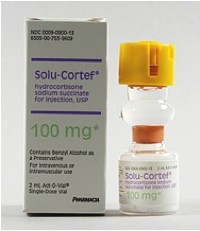
The specifc therapy for acute adrenal insufficiency is hydrocortisone, or Solu-Cortef:
Adult dose = 1 box, IM, IV, or IO.
Now, CT does not have any protocols so far regarding this medication. You can check out the protocols that RI, MA, NH, and NY have, though. (Those are all PDF downloads, BTW)

So, be ready for the change when it comes!

Sunday, December 11, 2011

Hypoglycemic patients, and refusal of transport.

It's a fairly typical ambulance run - you're called for "low blood sugar," and on arrival the wife points you to a sweaty, pale man who is shaking and speaking nonsense. The finger stick is "LO," but a box of D50 brings him around fairly quickly. You soon determine that he had administered his insulin just prior to lunch, but some distraction prevented him from eating on schedule.

No problem sir, just sign right here!
Probably not the best AMA form to be using...

For such a common practice, you would imagine that this would be a fairly safe encounter. Most EMS providers are careful to ensure that the patient will another person with them, that they eat a meal before the ambulance leaves, etc. Nonetheless, there are still a number of potential pitfalls that await the unwary paramedic.
Let me point out 5 important aspects of the "routine" hypoglycemic patient encounter:
  1. In general, this is a safe practice that patients are very satisfied with.
  2. Hypoglycemia due to oral hypoglycemics can be prolonged.
  3. Accidental ingestions of oral hypoglycemics by children, even if they are just suspected or possible, must always be transported, and admitted to the hospital.
  4. Hypoglycemia associated with long-acting insulin is also dangerous, and should also be transported.
  5. After a hypoglycemic episode, patients are especially vulnerable to a repeat episode, often showing symptoms at a higher glucose level.

Signing off patients is generally safe.

A number of studies, using a variety of methods and protocols, have shown that treating patients at home, and permitting them to refuse transport is a safe and popular practice. Studies conducted in the U.S. have more often used protocols that used an on-line medical control physician to talk with the paramedic, and often the patient as well. They also have used explicit inclusion criteria, and formal AMA paperwork. One study out of Buffalo used this inclusion check-off sheet:

Patients who met the criteria, and who had discussed refusing transport with the medical control physician, were given this form:

This level of documentation is very thorough, and likely prevents many potential lawsuits. The down-side of this elaborate protocol was that, over 3 years, less than 1/3 of the medics in that EMS system enrolled any patients, and only 36 patients total were enrolled.

Contrast this with one Canadian study, where only 34% of hypoglycemic patients ended up being transported. There was still a requirement for the paramedic to contact medical control, but there were no extensive inclusion criteria to follow. Instead, clinical discretion was expected of the medics. Perhaps that's why, in less than 1 year, 145 patients were found to decline transport - a far higher number than in Buffalo. Nonetheless, the practice was very safe, with recurrence of hypoglycemia happening just as often in the transported group as the refusals.

Even further from the U.S., both in protocol and in geography, is Finland. Researchers examined how well the protocols were working for them. Paramedics only needed to transport if patients "did not regain consciousness, were not able to eat after treatment, were pregnant, had type II diabetes treated with oral medication only or oral medication in addition to insulin, in case of insulin overdosage and variably in case the patient would be left alone or was a child (physician consultation)." In general, paramedics did not contact base physicians. Only 10% of patients ended up being transported!

Hypoglycemia due to sulfonylurea drugs must be transported.

Note that in the Finnish study, despite the willingness to leave many of the hypoglycemic patients at home, they required transport for patients who were taking oral hypoglycemics. Why was that?

Two reasons: duration of action, and underlying causes.

First, you must understand that not all oral diabetic medications are alike. There are five classes of oral medications: sulfonylureas, meglitinides, bigiuanides, alpha-glucosidase inhibitors, and the thiazoladinediones. While they all act in some way to prevent hyPERglycemia, only the first two classes are able to cause hyPOglycemia.

The most common sulfonylureas are glyburide, glimepiride, and glipizide, and they all have pretty long duration of action (24+ hours for both glyburide and glimeride). This means that if the patient accidentally took 2 or 3 pills extra today, 1 box of D50 and a PB&J sandwich may only temporarily bump the glucose, before the levels ground out again. This may not be a patient you can safely leave at home - they may have trouble for the next few hours or days!

Certainly, skipping a meal, or running for an hour longer may also promote hypoglycemia. These causes will be clear from the history. However, there are other causes of hypoglycemia associated with the oral medications, and the history may not be obvious.

Such causes are:
  • In patients who are undernourished or abuse alcohol
  • In patients with impaired renal or cardiac function or gastrointestinal disease
  • Concurrent therapy with salicylates, sulfonamides, gemfibrozil, or warfarin

These comorbidities often play a role in hypoglycemia. If you have a patient taking glyburide who has had an episode of hypoglycemia, realize that this may be just the "tip of the iceberg." We will end admitting a good chunk of these patients, so don't leave them at home. The little evidence we have about the safety of not transporting is not encouraging.

Speaking of the oral medications; this isn't strictly on-topic, but while we're on the topic...

The suspected ingestion of a sulfonylurea by a child gets transported.

Obviously, if a child is symptomatic from such an ingestion, they will be transported. The danger lays with the child whom the caretakers think may have taken a pill or two out of grandmother's purse. Despite normal glucose levels at the time of evaluation by EMS, patients have been known to develop hypoglycemia many hours after ingestion. This delayed effect seems to happen when children are given dextrose or food to prevent hypoglycemia, but it just puts it off for a few hours.

These are simple cases - they will all be admitted, even if there is just the suspicion of an ingestion.
"Kid and glyburide means ambulance ride."


Hypoglycemia associated with a long-acting insulin should be transported.


Lantus (insulin glargine) was approved by the FDA in 2000, and was the first insulin that was effective for 24 hours. This was a great therapeutic development, providing patients a more natural delivery of insulin. Levimer (insulin detemir) is the other commonly-used long acting insulin

Unfortunately, most of the good studies that looked at EMS "treat and release" protocols were conducted either before, or soon after, the release of Lantus. As a result, it is unclear if it would be safe to allow patients on these long acting insulins to refuse transport - we just don't have the data yet.

In the ED, these patients will usually be admitted, so don't think too hard about transporting!


Acute hypoglycemia predisposes patients to more recurrences.


Episodes of severe hypoglycemia seems to make it easier for a patient to have more episodes in the short term. Patients seem to lose both the ability to feel when their sugar is low, and a weaker capability to send out catecholamines to raise the sugar.

As a result, patients may have symptoms of hypoglycemia at a lower glucose level than before. If they developed an altered level of consciousness at a level of 55 mg/dL the first time, they may need to drop down to 35 mg/dL the second time.

For that reason, especially if patients decline transport to the hospital, they need to be educated that they should keep their glucose levels higher than usual for the next few days. In truth, it could take a few months of blood sugars in the 160-180 range to significantly improve their hypoglycemia awareness.

What do the Greater Bridgeport protocols say?

Glad you asked.

This protocol can't cover all the possibilities - we need an educated prehospital practitioner in the field to look at the scene, the patient, and get the history. But when you call in, you'll now have a much better idea of the things we're listening for, and the ways we can avoid the pitfalls.

Friday, December 2, 2011

ResQ-Pod 2: The story, and device, continues...

ResQ-Pod 2: Pod Harder.

 This study caught my eye caught my eye because the lead author is Stephen Smith, the author of one of the best ECG blogs out there. He also writes for other blogs, and at least one EMS ECG blog follows his work. Dr Smith is, in short, the man.

 The study also caught my eye because it uses a retooled version on the ResQPOD, now called the ResQGARD. I know a lot of EMS folk swear by the ResQPOD, but the recent evidence has not proven its value. So, it's interesting to see "part 2" of the ResQPOD saga.
Because Part 1 worked out so well.

The ResQGARD works on the same general physiologic principle as the ResQPOD. It allows for normal, unimpeded exhalation, and does not provide any PEEP. During inhalation, however, it slightly increases the force required to draw in a breath. The actively expanding thorax normally acts as a sort of "suction" to also pull blood up from the belly, but with this added resistance to the air inflow, this "suction" effect is magnified.

And there is some animal and human data to back up the claims for usefulness in treating hypotension.
Red means more blood, I guess.

This device is evidently "cleared" by the FDA for treating "low blood circulation," and various studies have shown an ability to raise the blood pressure in, for example, blood-donors, or in other models of hypovolemia or hemorrhage. You can check out some background device in this article from Journal of Special Operations Medicine, the coolest journal that you aren't reading yet.
"Oh, is that JEMS you're reading? That's cute."

This trial had two parts. In the first part, the device was tested in a randomized, controlled, and blinded fashion in the emergency department for patients with hypotension due to various causes. The primary endpoint was the maximum change in SBP over the first 10 minutes after placement of the device. They enrolled 47 hypotensive patients. These patients ended up with diagnoses of dehydration, sepsis, or hemorrhage most of the time. You can see the average change in SBP in the table below, broken down by the cause of the hypotension.

When they looked at the overall results, they found that patients who had the real device had an average rise in SBP of 12.9 mmHg, while patients who got the sham device only had a rise of 5.9 mmHg, a difference of 7 mmHg. They tell us that the difference is statistically significant.

In the second part, the device was used by EMS, with no control therapy, in an unblinded manner. It's not much of a study, and they were really only looking at "feasability" of using the device by prehospital providers. They also had 47 patients in this arm of the study, and they determined that, yes, it was feasible to use, and that patients tolerated the device well.

It's hard to interpret the data on the change in blood pressure, etc., in part 2, since, as we saw in the results of the first part, the average blood pressure tended to go up with or without the device. The bar graph below shows that the pressure came up to a statistically significant degree, but it can't tell us if this was better than doin' nuthin'.

So, what can we take from this paper?

In the end, not much. Let me list the reasons why:
  1. The difference in SBP is statistically significant, but unclear if clinically significant.
  2. Some of the causes for hypotension have established, beneficial treatments.
  3. Some of the causes require no treatment, and improve on their own.
  4. The majority of the literature supporting the use of this device is written, in part, by the inventor of the device.
The average difference in systolic blood pressures was 7 mmHg, which is pretty small. How much do you care about raising the pressure by 7 points? In addition, it looks like almost everyone's pressure went up somewhat, with or without the device. Remember that 7 represents the average difference - some patients didn't improve much with the device, and some people had a big jump in pressure with the fake device!

In another article about the ResQGARD (or ITD-7) written by Smith, the authors present this table:
"Treatable" ≠ "Proven effective for"

Let's look at the causes they list. First off, heat stroke is about heat load and mental status - the primary treatment is cooling, and hypovolemia is usually not a significant component of the problem.

The proper treatment of true dehydration is, well, hydration. As for sepsis, while hypotension is a manifestation of the problem, there is a large amount of evidence that large amounts of IV fluids, delivered rapidly, saves lives. Simply raising the pressure through other means is not appropriate.

Regarding hemorrhage, there is a thicket of controversy about optimum treatment. While much of current practice emphasizes normalizing the blood pressure, a lot of evidence suggests that "permissive hypotension" may be the best (non-)treatment. Where the ResQGARD falls in this area is not clear at all.

Lastly, orthostatic hypotension is usually transient, and requires no intensive therapy; e.g. juice & cookies after blood donation.

Let me speak of the appearance of bias in the studies supporting the ResQGARD. The inventor of the device is Keith Lurie, a cardiologist. I have no doubt that he has aspirations to advance medical science and save lives. Unfortunately, as the inventor of the device in question, and the owner of the company that sells them, he has a vested interest in selling the device. And, while it's not the most expensive medical device out there, it costs real money.

Look at the references listed in the paper. Of the 23 studies that Smith et al. provide as references, 20 had Dr. Lurie as a co-author. That's a real conflict of interest. By way of example, check out this intervew that appeared with Dr. Lurie in an EMS blog. He had an interesting take in the failure of the ResQPOD to show an effect in the ROC trial.

Interviewer: "I think many of us who have been following the ResQPOD were surprised by the recent announcement by the National Institute of Health that the ROC PRIMED trial was stopping enrollment. ...  Considering that the ROC PRIMED trial was a prospective, multi-centered, randomized clinical trial with large enrollment, are you concerned about these results?

Dr. Lurie: "To directly answer your question, I am not concerned with the results, nor am I surprised."

To sum up: The ResQGARD appears to have a statically significant effect in hypotensive patients, but the clinical effect, as well as the appropriateness of this therapy, are unclear. I don't think any EMS service should be stocking up on these yet.